Online Course
Diagnostic Endoscopy
– An Immersive Guide –
Presented by Dr. Jacqueline Whittemore
DVM, PhD, DACVIM
Please Note:
The course needs to be completed within six months
of the registration date, after which it will expire.
Minimally-invasive Scoping
This course is designed to provide in-depth training in small animal endoscopy with a focus on techniques used by small animal internists.
The successful participant will learn the fundamentals of flexible and rigid scope anatomy, handling and cleaning; laser physics, handling and safety; cystoscopy, rhinoscopy, bronchoscopy, upper and lower gastrointestinal endoscopy, and laparoscopy.
Course Structure & Requirements
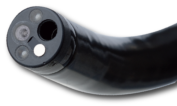
Module 1: The Flexible Endoscope
This module contains 4 parts:
- Anatomy of the flexible endoscope: Basic scope function and anatomy will be reviewed, with special attention given to selection considerations and troubleshooting.
- Other instrumentation: Components of the endoscope tower, their function, and considerations in use will be presented. This module will also present and discuss differing instruments available for use with the flexible endoscope.
- Handling and manipulation: In this module, appropriate scope handling and manipulation will be reviewed. The trifecta of patient, tower, and endoscopist positioning – and its impact on procedure outcome – will also be discussed.
- Cleaning the flexible endoscope: In this module, the complex task of scope disassembly and cleaning will be broken down into easy to understand and manage steps. Cleaning of flexible instruments will also be reviewed.
Module 2: The Rigid Endoscope
This module contains 2 parts:
- Anatomy of the rigid endoscope and other instrumentation: Basic scope function and anatomy of the rigid endoscope, variations in scope tower components, and rigid endoscope instruments will be discussed.
- Handling, manipulation, and cleaning: Essentials of successful rigid scope handling and common areas of disaster are broken down, followed by a step by step review of cleaning of rigid equipment.
Module 3: Ancillary Equipment
This module contains 2 parts:
- Laser physics, safety and handling: Fundamentals of laser design and physics are discussed with focus on clinical application and laser selection. Finally, core tenets of laser handling, safety, and maintenance are reviewed.
- Ancillary equipment: This module reviews supplemental equipment commonly used for laparoscopic and thoracoscopic procedures with a focus on ideal technique.
Module 4: Cystoscopy
This module contains 4 parts:
- Overview and transurethral cystoscopy in the female: General advantages, disadvantages and indications for cystoscopy are reviewed, after which the particulars of transurethral cystoscopy in the female is described.
- Transurethral cystoscopy in the male, prepubic cystoscopy: Performance of cystoscopy in the male dog will be reviewed with attention given to use of a standard transurethral versus a perineal approach. Non-urethral approaches for cystoscopy will also be described.
- Special procedures – urinary anomalies: Urethral bulking and correction of ectopic ureters, ureteroceles, and vestibulovaginal remnants are discussed. Indications, advantages, disadvantages, and particulars of procedure performance are covered.
- Special procedures – acquired conditions: Laser lithotripsy, urethral and ureteral stent placement, and sclerotherapy for benign renal hematuria are reviewed. Keys in patient selection and positioning are covered in addition to indications, advantages, disadvantages, and particulars of procedure performance.
Module 5: Rhinoscopy
This module contains 4 parts:
- Overview and retrograde rhinoscopy: General advantages, disadvantages and indications for rhinoscopy are reviewed, after which the particulars of retrograde rhinoscopy is described.
- Antegrade rhinoscopy: Navigation of the dorsal, middle and ventral meatuses during antegrade rhinoscopy will be explained, as well discussion of keys to procedure performance and collection of diagnostic samples.
- Special rhinoscopy procedures: Nasopharyngeal stenosis correction, canaliculocele omentalization, and sinusotomy tube placement are reviewed. Indications, advantages, disadvantages and particulars of procedure performance are covered.
- Special rhinoscopy procedures: A detailed breakdown of sinonasal aspergillosis management techniques is presented, followed by a review of cases with comorbid nasal diseases and key tenets for successful procedural outcome.
Module 6: Bronchoscopy
This module contains 3 parts:
- Overview and tracheoscopy: General advantages, disadvantages, indications and complications of bronchoscopy are discussed, with particular attention given to patient oxygenation. Tracheoscopy technique and grading of tracheal collapse then are reviewed.
- Bronchoscopy: Oxygenation during bronchial intubation, navigation and evaluation of the bronchi, and the impact of rotation control are dissected.
- Bronchoscopic procedures: Bronchoalveolar lavage (BAL), the keystone bronchoscopic procedure, is thoroughly reviewed. Discussion of BAL is followed by details on airway biopsy, tracheal mass resection and foreign body retrieval, as well as a detailed tutorial on tracheal stenting.
Module 7: Esophagogastroduodenoscopy
This module contains 5 parts:
- Overview and basic technique: General indications, advantages, disadvantages, and complications of upper gastrointestinal endoscopy are discussed, followed by details on exploring the esophagus, stomach and duodenum.
- Diagnostic sampling: Criteria for ideal forceps selection under differing biopsy conditions are briefly reviewed. This is followed by a discussion of the differing techniques for biopsy collection, important sample handling considerations, and the impact of staining on diagnostic capability.
- Foreign body retrieval: Location of foreign bodies, risk factors, and impact of timing of retrieval on outcome are broken down, followed by discussion of techniques with examples given of retrieval of differing types and locations of objects.
- Esophageal stricture correction: Theoretical advantages and disadvantages of bougienage and balloon dilation are contrasted. Management of initial esophageal strictures, recurrent strictures, and panesophageal strictures is presented, with cases used to highlight key factors in procedure performance.
- Feeding tube placement: Placement of homemade gastrostomy tubes, one-step buttons, and replacement gastrostomy tubes is described and illustrated using cases. Jejunostomy, nasojejunostomy and esophagojejunostomy tubes are also discussed. Particular attention is given to contraindications and complications of tube placement, as well as strategies to mitigate risk of their occurrence.
Module 8: Colonoscopy, plus
This module contains 3 parts:
- Overview and patient preparation: General indications, advantages, disadvantages, and complications of colonoscopy and ileoscopy are reviewed. Equipment necessary for colonoscopy and proctoscopy is reviewed. Keys to achieve optimal preparation of the lower bowel are discussed, as are details necessary for successful management of catastrophic complications.
- Basic technique: Diagnostic colonoscopy and ileoscopy are described, with particular attention given to discussion of common navigational challenges, anatomic factors that contribute to them, and adjustments in technique to maximize success. Sampling techniques unique to the descending colon are demonstrated.
- Capsule endoscopy: General indications, limitations and complications of capsule endoscopy are reviewed. The specifications of different types of endoscopy capsules are broken down, along with their advantages and disadvantages, to allow selection of the capsule endoscopy system best suited to the endoscopist’s needs. Finally, current literature on capsule endoscopy use in dogs is discussed.
Module 9: Laparoscopy
This module contains 5 parts:
- Basic overview and restraint: Indications and contraindications for laparoscopy, necessary instrumentation, and important anesthetic considerations are presented.
- Patient positioning and entry techniques: Patient preparation, commonly used positions, and types of laparoscopic approach will be presented with special attention paid to selecting the best approach for each procedure.
- Laparoscopic explore: First entry and placement of cannulae, along with complications, are reviewed. Several examples of general exploration are presented, followed by a discussion of closure steps and tips/tricks for laparoscopy.
- Laparoscopic procedures: Biopsy of liver, pancreas, and spleen are presented. Application of Gelfoam and performance of cholecystocentesis are also discussed.
- Laparoscopic procedures, part 2: Performance of renal biopsy and full-thickness intestinal biopsy are discussed, followed by review of jejunostomy feeding tube placement. The module concludes with a discussion of tips and tricks.
PRICING
VETERINARIAN
$1,000.00 USD
VETERINARIAN TECHNICIAN
$500.00 USD
RESIDENTS
$750.00 USD
Credits: 20 CE Hours
Available Languages: English (US)
Individuals living in disadvantaged or developing countries (see Research4Life) are candidates for full or partial reduction in registration fees for this course.
Eligible individuals should contact the course administrator prior to registration regarding this potential opportunity.
Terms and Conditions apply.
An Integrated Live Course combining online training with a hands-on, immersive laboratory experience is also available. Click here for more information.
FACULTY:
DR. JACQUELINE WHITTEMORE
DVM, PhD, DACVIM
Lead Instructor
The Karl Storz Diagnostic Endoscopy course was awesome! Jackie is such an engaging teacher and very approachable. I learned some great tricks and realized I had been doing some things wrong for over a decade!
– Amber
”
And I am long (long, long) overdue for a huge thank you for the course.
It was incredible, amazing, worthwhile, and 100% re-inspiring for me as an internist.
– Online course registrant
”
Just a note, Dr. Whittemore, to let you know how much I admire your dedication to sharing your amazing talents. I have learned so much from you and your associates. I have done four practice upper GI procedures on two employees’ animals needing routine procedures and two rescued animals being neutered. All four allowed me to advance into the duodenum and get some much needed practice. I am determined to build the confidence to put my UT skills to work. Also, I am cleaning this expensive equipment properly, thanks to your CE course. Again, your enthusiasm makes us old guys want to keep learning. Thanks. – Dr. Larry Hendricks
”
I just wanted to give you a shout-out today… I’ve literally used my endoscope twice now in one week. Once to confirm the presence of a pyloric outflow obstruction that led to a gastrotomy/enterotomy. And today with a chronic vomiting young boxer, with whom I was able to pass into the duodenum on the first fricking try! It ended up having a focal area of gastric ulceration. I literally hooted and hollered when I navigated into the duodenum. Thank you! See you in August.
– Brian Dickinson
”
I have been progressing through the modules and am extremely embarrassed to say I performed the worst on the cystoscopy final! You have done an amazing job with this course and I really think it should be mandatory for all internal medicine residents in their first year and then repeated in their final year. I would have been so much more effective had I taken this training at the beginning of my residency (although you were probably still in high school then).
– Sheri J Ross
”